Segmental Diagnostic Findings
with Testicular Cancer
Draw clear conclusions and act immediately
Cancer diagnostics can be called a stepchild of energy medicine. This is based, among other things, on the fact that every cancer type is different and specific type diagnoses are hardly possible. You can also say that for the patient’s well [being] the individuality of the diagnoses is rarely as distinctive as with cancer. From a spiritual perspective, you can establish that with the development of cancer the soul empties all its negative contents and its repressed traumas and stresses out into the subconscious, and inserts them into this disease.
In some cases it is possible to raise a suspicion by means of the Carcinosinum nosode or specific cancer nosodes, however this cannot be considered as an understandable rule because the specificity is too low. Therefore a case should be described in which without understanding it, a cancer diagnosis finding was registered by means of the AMSAT-HC® device. Three months later two distinctive cancer types could be found. The question arises: Could you have already expressed the specific suspicion in the early stage?
The 36 year old patient felt a knot in his right testicle three months after his segmental diagnostic examination. Because he was HIV-positive for some years and was treated accordingly, it naturally frightened him very much. Then examinations like sonography, laboratory work including tumor markers, explorative biopsy, operation and histology resulted in the following diagnosis (quoted from the physician’s letter):
“Seminoma of the left testicle
pT1 with rete testis infiltration, pNX, pMX, V0, L0, R0
AJCC Stage 1A, clinical stage I “intermediate risk”
Seminoma and embryonal cell carcinoma of the right testicle.
pT2, pNX, pMX, L1, V1, R0
AJCC Stage IB, clinical stage I “high risk”
Cell necrosis inguinal ablation right testis and tumor enucleation on the left.
28.10.2009
HIV CDC stage IA”
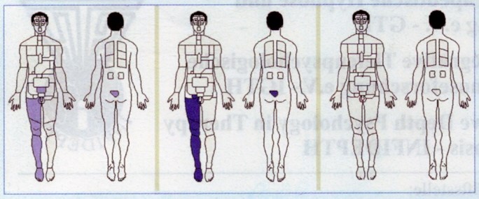
Undoubtedly this is a problematic and unfortunate development. Now we look at the findings raised by the segmental diagnostics. The evaluation occurs according to three criteria: Base (= Function), Sol-Gel (= solid tissue) and Risk (= sum of both). You see the results on the “phantom images” with topographical representation of the organs (worst tested short measurement) in Figures 1a through 1c. In all three images you recognize pathological findings of the right testicle. The representation of the right leg speaks for a lymph drainage disorder and consequently a burden on the right side inguinal lymph nodes.

Figure 1
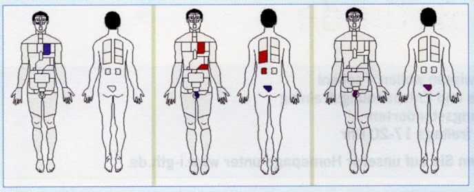
If you pick out the results of the best short measurement, this yields the Figures 2a (Base), 2b (Sol-Gel) and 2c (Risk).

Figure 2
Analysis of the function in Figure 2a shows a hypo-state of the heart, most likely psychological. In Figure 2b (solid tissue) you see a Gel-state of the left testicle. The Sol-state of the pancreas and heart most likely shows up as a result of the connection to the HIV basic illness. In Figure 2c (Risk) the right testicle again steps into the foreground.
In retrospect you can capture the suspicion of serious pathological findings in the testicles three months before the clinical diagnosis of cancer illness. However no suspicious diagnosis occurred concerning a malignant development except for an optimization of medication therapy and the recommendation of a visit to a urologist. This was based on the previously mentioned far too self critical basic position of an insufficient specificity of an energy medicine examination result.
Conclusion
The findings shown here should give us courage to trust the results, to draw clear conclusions from it, and to press for an immediate clinical clarification of localized pathologies. The fear of an HIV+ patient of further illnesses is understandable and therefore the effort to spare him should not be an obstacle. If the patient had immediately gone for a sonograph examination, the diagnosis could have been put to his advantage earlier.
 An Exclusive Translated Article for Members
From THE BRIDGE Newsletter of OIRF
Published April 2010
From an article in CO’MED, No. 1, 2010
Machine Translation by SYSTRAN, Lernout & Hauspie, LogoMedia & Promt
Translation & redaction by: Carolyn L. Winsor, OIRF
© Copyright 2009, Dr. med. Manfred Doepp, Prien, Germany


